Doppler ultrasound for dairy cattle reproduction gives veterinarians an additional way to evaluate reproductive status in the field. Like conventional rectal ultrasound, the examination is performed to assess reproductive structures. The important difference is that Doppler imaging can display blood flow within blood vessels. This added information can support a more complete assessment of the ovaries, uterus, follicles, and corpus luteum.

For dairy herd management, reproductive decisions often need to be made promptly and with the best available information. Doppler ultrasonography does not replace veterinary judgment or conventional ultrasound. Instead, it adds a blood-flow perspective that may help clarify whether a reproductive structure is functioning as expected.
What Doppler imaging adds to reproductive examinations
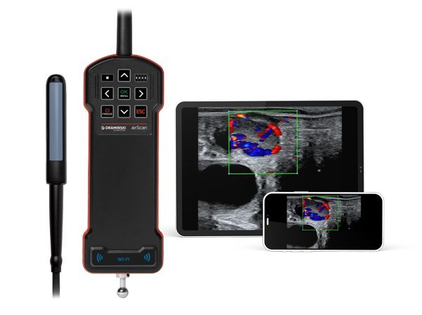
Conventional ultrasound helps visualize reproductive anatomy. Doppler imaging extends this examination by assessing vascular flow. In dairy cattle reproduction, this can help veterinarians evaluate the condition of the ovaries and uterus while also considering the vascular activity associated with key reproductive structures.
This capability is particularly relevant when examining follicles and the corpus luteum (CL). A follicle may be evaluated not only by its visible appearance but also by whether it appears active and likely to ovulate or is already undergoing atresia. Such observations can help reduce uncertainty when planning the next steps in a breeding program.
Assessing corpus luteum function
The corpus luteum plays an essential role in maintaining pregnancy. With Doppler ultrasonography, a veterinarian can assess blood flow associated with the CL and evaluate its function before an obvious change in size occurs. This may be useful when investigating an insemination that has not resulted in the expected outcome.
The source information also notes that Doppler evaluation may assist with failed insemination assessments, including situations before day 28 after breeding. Earlier recognition of an unsuccessful pregnancy can allow a herd to move more quickly toward an appropriate rebreeding decision under veterinary guidance.
Supporting pregnancy and embryo transfer decisions
Doppler ultrasound can contribute to earlier evaluation of pregnancy status or the absence of pregnancy. The value is not limited to confirmation alone: blood-flow information may provide additional context when interpreting reproductive findings and planning herd management actions.
In embryo transfer programs, recipient selection is another potential application. A well-vascularized corpus luteum is associated in the source material with improved conditions for embryo implantation. The source also states that CL perfusion can be a stronger predictor than CL size or progesterone level when selecting recipients. This makes blood-flow assessment a practical consideration for veterinary professionals working with embryo transfer protocols.

Potential management value for dairy herds
More informed reproductive examinations can support better herd-level decisions. When Doppler findings are interpreted by a veterinarian alongside clinical history and standard ultrasound results, they may help avoid unnecessary inseminations and the associated semen use.
- Earlier identification of failed pregnancies may support quicker rebreeding decisions.
- Follicle and CL assessment may help refine the use of hormonal protocols.
- Recipient selection for embryo transfer can be based on more than visible CL size alone.
- Reproductive performance can be reviewed with additional information when evaluating animals for breeding programs.
These considerations are especially meaningful in larger dairy herds, where breeding timing, calving interval, successful insemination, and embryo transfer outcomes all influence operational planning. Doppler ultrasonography provides information that can help veterinarians and herd managers make decisions with fewer assumptions.
Is Doppler ultrasound worthwhile in dairy cattle reproduction?
The value of Doppler ultrasound depends on how it is used within a herd’s veterinary and reproductive management process. Its central advantage is the ability to assess blood flow in addition to anatomical structures. This can strengthen the evaluation of ovarian activity, corpus luteum function, pregnancy status, and embryo transfer recipients.
For practices and dairy operations seeking a more detailed reproductive assessment, Doppler ultrasonography is a relevant tool for modern cattle reproduction. It supports a more informed approach to breeding decisions while keeping the veterinarian’s clinical interpretation at the center of the process.